
FASCIA, HERNIAS, AND OTHER INJURIES
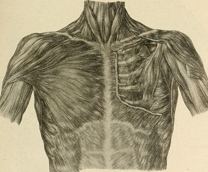
“Interest in elucidating the etiology of hernias has encouraged countless studies of musculoaponeurotic structures in individuals with and without hernias. Studies of hernia patients have firmly demonstrated a correlation between hernias and collagen alterations in their fascia.”
The picture above (as well as the quote) comes from Dr John Deaver’s 1917 textbook, The Breast: its Anomalies, its Diseases, and their Treatment, and shows a great example of the linea alba or “white line”.
The linea alba is an APONEUROSES that runs from the bottom of the chest (xiphoid) to the union of the two pubic bones (pubic symphisis), separating the right and left rectus abdominus muscles (the muscles known for their “six pack” appearance in thin individuals). Diastais Recti is the condition where to varying degrees, the center line tears and splits apart, allowing underlying musculature to “pooch” through (WebMD says that “about two-thirds of pregnant women have it“). That’s right; it’s yet another one of the numerous problems that tend to afflict women more than men.
The quote at the top of the page comes from a Brazilian study titled Collagen I and III in Women with Diastasis Recti that was published in last month’s issue of the journal, Clinics. In this study, the authors biopsied FASCIA that was collected from the linea alba — a common area for women to get hernias (HERE is another that may be just as common). Some women had hernias, compared to an equal number of women without. Below are some of the CHERRY-PICKED findings.
As a constituent of the abdominal fascia and aponeuroses, including the linea alba, collagen has the important structural role of providing support and resistance to the abdominal wall against intra-abdominal pressure. Type I collagen is the most abundant form; it is the main component of aponeuroses, tendons, and mature scar tissue, and its main function is to provide resistance to tensile stress.
Type III collagen corresponds to what was classically described as reticular fiber, and its function is to provide support to expandable structures. Type III collagen levels also increase during the early stages of wound repair. Studies have shown that patients with hernias of various etiologies have lower type I and III collagen levels than control cadavers with no history of hernia, highlighting the importance of collagen in the body’s support structures.
This is interesting in that patients with various sorts of hernias (abdominal, inguinal, hiatal, etc, etc,) have fascia that is actually different than normal — different than people without hernias. That’s great to know but my question is what can be done to change this? What can be done to help those women (or men) dealing with Diastasis Recti? One of the more interesting things that these authors said was that according to previous research, this problem can go away on its own — a fact verified in this study’s conclusions.
Which begs the question; are there specific things people could be doing to prevent or even reverse DR?As is the case with the solution to most health issues, solving a separated abdomen will likely require a multi-faceted approach. Furthermore, when done correctly, at the very worst, a good protocol will not worsen your problem but could instead benefit you in a variety of ways — particularly if you are a person who is “INJURY-PRONE“.
- FIRST: Get rid of inflammation in your life. Once you realize that even WEIGHT GAIN is considered an “INFLAMMATORY PROBLEM,” this bullet starts making more sense. While diet is certainly important for reducing inflammation (HERE is my favorite method of eating, with THIS coming in a close second; particularly if you need to lose a significant amount of weight in the process), there are any number of things you might be exposing yourself to that have the potential to drive inflammation.
- SECONDLY: Increase your intake of COLLAGEN. While there are certain genetic factors that you cannot change about the way your body deals with collagen (HERE and HERE are examples), you can certainly change the EPIGENETIC FACTORS which are actually a bigger deal for most people anyway. Although there are numerous excellent collagen products on the market (as well as numerous crappy products), I recommend good, old-fashioned, bone broth. In my humble opinion, the secret to high quality bone broth that you will actually look forward to consuming, is to essentially make soup broth (onions, ANTI-INFLAMMATORY SPICES, etc, etc). Just stay away from the MSG-BASED bullions and make sure you are simmering and not boiling. If you’re still not sure, not only are there numerous recipes on YouTube, ask the elderly women in your circle of friends or your church.
- THIRDLY: This is the place everyone wants to start — abdominal strengthening exercises. And funny enough, most people want to move straight into the single worse exercise for contributing to DR, SITUPS. A gentle overall CORE-STRENGTHENING PROTOCOL (mine is probably a bit too stringent for this use) is almost always a good thing, with dozens of sites on the web devoted to the topic. And although some of them are pay-to-play, free information abounds. As a man, I can’t really vouch for any of them, but after looking at many, they all seem to be rather similar. Just Google “healing a diastasis recti naturally,” see what comes up, and start formulating your own personalized plan.
For those of you that DR is just the tip of the iceberg of chronic problems, I have a bit more comprehensive generic protocol for regaining your health (HERE). While it’s certainly not going to solve everything for everyone, it’s going to help you start thinking differently about certain health-related subjects (for instance, GUT HEALTH). If you are enjoying what you are learning on our site, be sure to help others. Forward a link or simply like, share, or follow on FACEBOOK as it’s a fantastic way to reach those you love and care about most.